| Citation: |
ZHANG Nan, JIN Xuanyi, LI Guangyuan, MA Chunyan. Value of different patterns of complete left bundle branch block evaluated by two-dimensional speckle tracing echocardiography in predicting acute response of patients with cardiac resynchronization therapy[J]. Journal of Clinical Medicine in Practice, 2023, 27(7): 1-5, 24. DOI: 10.7619/jcmp.20223548

|
To explore the clinical value of different patterns of complete left bundle branch block (CLBBB) evaluated by two-dimensional speckle tracing echocardiography (2D-STE) in predicting acute response of chronic congestive heart failure (CHF) patients with cardiac resynchronization therapy (CRT).
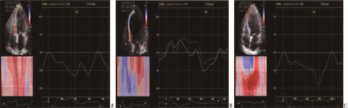
A total of 36 patients with CHF and CLBBB by CRT were selected and conducted with echocardiography examination in the ON and OFF states of CRT, and the increase of left ventricular ejection fraction (LVEF) ≥ 5% at ON state of CRT was defined as responsive, while the increase of LVEF < 5% was defined as unresponsive. According to the longitudinal time-strain curve types of left ventricular posterior interventricular septum, CLBBB was divided into type Ⅰ, type Ⅱ and type Ⅲ. Patients with type Ⅰ and type Ⅱ were set as study group 1, patients with type Ⅲ were set as study group 2, and the conventional ultrasound parameters, systolic function parameters and systolic asynchrony parameters were detected at the OFF state of CRT.
Among the 36 patients, there were 29 cases (80.56%) with response and 7 cases (19.44%) without response; there were 20 patients with type Ⅰ CLBBB, 4 patients with type Ⅱ, and 12 patients with type Ⅲ. There were 22 patients with effective response to acute response of CRT in the study group 1, and the effective rate of response was 91.67% (22/24); in the study group 2, 7 patients had effective response to acute response of CRT, and the effective rate of response was 58.3% (7/12); the effective rate of response to acute response of CRT in the study group 1 was significantly higher than that in the study group 2 (P < 0.05). There were no significant differences in left ventricular diameter, left ventricular systolic function, left ventricular diastolic function and ventricular asynchrony parameters between the two groups (P>0.05). The overall longitudinal peak strain of the interventricular septum and the overall longitudinal peak strain of the lateral wall in the study group 1 were significantly lower than those in the study group 2 (P < 0.05). The standard deviation of the peak time of left ventricular strain at 18 segments in the study group 1 was significantly greater than that in the study group 2 (P < 0.05).
There are differences in response efficiency of acute response among different types of CLBBB; the response efficiency of acute response in type Ⅰ and Ⅱ CLBBB is better than that of type Ⅲ CLBBB; the function of interventricular septum and left ventricular sidewall in type Ⅰ and Ⅱ CLBBB is better than that of type Ⅲ CLBBB; the left ventricular systolic asynchrony of type Ⅰ and Ⅱ CLBBB is more significant than that of type Ⅲ CLBBB.
| [1] |
EUROPEAN SOCIETY OF CARDIOLOGY (ESC), EUROPEAN HEART RHYTHM ASSOCIATION (EHRA), BRIGNOLE M, et al. 2013 ESC guidelines on cardiac pacing and cardiac resynchronization therapy: the task force on cardiac pacing and resynchronization therapy of the European Society of Cardiology (ESC). Developed in collaboration with the European Heart Rhythm Association (EHRA)[J]. Europace, 2013, 15(8): 1070-1118. doi: 10.1093/europace/eut206
|
| [2] |
WANG M, XU Y, WANG S, et al. Predictive value of electrocardiographic markers in children with dilated cardiomyopathy[J]. Front Pediatr, 2022, 10: 917730. doi: 10.3389/fped.2022.917730
|
| [3] |
刘广志. 心源性卒中的诊治[J]. 中华脑血管病杂志: 电子版, 2021, 15(4): 275-275. https://www.cnki.com.cn/Article/CJFDTOTAL-ZHND202104018.htm
|
| [4] |
睢勇, 孙慧, 幺传为, 等. 慢性心力衰竭患者预后不良的血清学预测指标研究[J]. 实用临床医药杂志, 2021, 25(21): 52-57. doi: 10.7619/jcmp.20212165
|
| [5] |
OUALI S, KACEM S, GRIBAA R, et al. Successful pregnancies after transvenous cardiac resynchronization therapy in a woman with congenitally corrected transposition of the great arteries[J]. Egypt Heart J, 2017, 69(3): 219-222. doi: 10.1016/j.ehj.2017.05.002
|
| [6] |
ZHANG W W, HUANG J J, QI Y D, et al. Cardiac resynchronization therapy by left bundle branch area pacing in patients with heart failure and left bundle branch block[J]. Heart Rhythm, 2019, 16(12): 1783-1790. doi: 10.1016/j.hrthm.2019.09.006
|
| [7] |
EZZEDDINE F M, SALIBA A N, JAIN V, et al. Outcomes of cardiac resynchronization therapy in patients with chemotherapy-induced cardiomyopathy[J]. Pacing Clin Electrophysiol, 2021, 44(4): 625-632. doi: 10.1111/pace.14196
|
| [8] |
TUNUGUNTLA H P, PURI K, DENFIELD S W. Management of advanced heart failure in children with cancer therapy-related cardiac dysfunction[J]. Children (Basel), 2021, 8(10): 872.
|
| [9] |
FUDIM M, DALGAARD F, AL-KHATIB S M, et al. Future research prioritization in cardiac resynchronization therapy[J]. Am Heart J, 2020, 223: 48-58. doi: 10.1016/j.ahj.2020.02.011
|
| [10] |
HENRY R, DOOKIE T, PRIMUS E. A comparative survey of pacemaker implantation in Trinidad and Tobago in 2005 and 2009[J]. West Indian Med J, 2014, 63(5): 474-478.
|
| [11] |
MELGAARD J, VAN DAM P M, SOMMER A, et al. Non-invasive estimation of QLV from the standard 12-lead ECG in patients with left bundle branch block[J]. Front Physiol, 2022, 13: 939240. doi: 10.3389/fphys.2022.939240
|
| [12] |
OWASHI K, TACONNÉ M, COURTIAL N, et al. Desynchronization strain patterns and contractility in left bundle branch block through computer model simulation[J]. J Cardiovasc Dev Dis, 2022, 9(2): 53. doi: 10.3390/jcdd9020053
|
| [13] |
中华医学会超声医学分会超声心动图学组, 中国医师协会心血管分会超声心动图专业委员会. 超声心动图评估心脏收缩和舒张功能临床应用指南[J]. 中华超声影像学杂志, 2020, 29(6): 461-477. doi: 10.3760/cma.j.cn131148-20200227-00115
|
| [14] |
SHOMAN K A, ELDAMANHORY H M, FAKHRY E E, et al. Role of Strauss ECG criteria as predictor of response in patients undergoing cardiac resynchronization therapy[J]. Egypt Heart J, 2022, 74(1): 69. doi: 10.1186/s43044-022-00308-3
|
| [15] |
CORTEVILLE B, DE POOTER J, DE BACKER T, et al. The electrocardiographic characteristics of septal flash in patients with left bundle branch block[J]. Europace, 2017, 19(1): 103-109.
|
| [16] |
ANDERSON K P. Left bundle branch block and the evolving role of QRS morphology in selection of patients for cardiac resynchronization[J]. J Interv Card Electrophysiol, 2018, 52(3): 353-374. doi: 10.1007/s10840-018-0426-z
|
| [17] |
ROSALIA L, OZTURK C, SHOAR S, et al. Device-based solutions to improve cardiac physiology and hemodynamics in heart failure with preserved ejection fraction[J]. JACC Basic Transl Sci, 2021, 6(9/10): 772-795.
|
| [18] |
CLELAND J G F, BRISTOW M R, FREEMANTLE N, et al. The effect of cardiac resynchronization without a defibrillator on morbidity and mortality: an individual patient data meta-analysis of COMPANION and CARE-HF[J]. Eur J Heart Fail, 2022, 24(6): 1080-1090. doi: 10.1002/ejhf.2524
|
| [19] |
LAU C, ELSHIBLY M M M, KANAGALA P, et al. The role of cardiac magnetic resonance imaging in the assessment of heart failure with preserved ejection fraction[J]. Front Cardiovasc Med, 2022, 9: 922398. doi: 10.3389/fcvm.2022.922398
|
| [20] |
GHOSSEIN M A, VAN STIPDONK A M, PRINZEN F W, et al. Vectorcardiographic QRS area as a predictor of response to cardiac resynchronization therapy[J]. J Geriatr Cardiol, 2022, 19(1): 9-20.
|
| [21] |
ĪZGI T N, BARUTCU ATAŞ D, ATAŞ H, et al. Prediction of subclinical left ventricular dysfunction by speckle-tracking echocardiography in patients with anti-neutrophil cytoplasmic antibody: associated vasculitis[J]. Arch Rheumatol, 2022, 37(1): 129-135. doi: 10.46497/ArchRheumatol.2022.8916
|
| [22] |
AL SAIKHAN L, PARK C, HUGHES A D. Reproducibility of left ventricular dyssynchrony indices by three-dimensional speckle-tracking echocardiography: the impact of sub-optimal image quality[J]. Front Cardiovasc Med, 2019, 6: 149. doi: 10.3389/fcvm.2019.00149
|
| [23] |
DURAL M, VAN STIPDONK A M W, SALDEN F C W M, et al. Association of ECG characteristics with clinical and echocardiographic outcome to CRT in a non-LBBB patient population[J]. J Interv Card Electrophysiol, 2021, 62(1): 9-19. doi: 10.1007/s10840-020-00866-z
|
| [24] |
RISUM N, KISSLO J, WAGNER G. Cardiac resynchronization therapy: identifying an activation delay by regional strain analysis[J]. J Electrocardiol, 2015, 48(5): 779-782. doi: 10.1016/j.jelectrocard.2015.07.020
|
| [25] |
RISUM N, TAYAL B, HANSEN T F, et al. Identification of typical left bundle branch block contraction by strain echocardiography is additive to electrocardiography in prediction of long-term outcome after cardiac resynchronization therapy[J]. J Am Coll Cardiol, 2015, 66(6): 631-641. doi: 10.1016/j.jacc.2015.06.020
|
| [26] |
VILLEGAS-MARTINEZ M, KROGH M R, ANDERSEN Ø S, et al. Tracking early systolic motion for assessing acute response to cardiac resynchronization therapy in real time[J]. Front Physiol, 2022, 13: 903784. doi: 10.3389/fphys.2022.903784
|
| 1. |
王磊,尤菲,张锋,吴冠吉,刘树文,马前锋. 老年心力衰竭患者应用心脏再同步化治疗后无应答的影响因素. 国际医药卫生导报. 2024(08): 1248-1252 .

| |
| 2. |
刘艳,陈小春,郑建民. 不同类型完全性左束支传导阻滞患者心室功能与心室内收缩同步性的差异. 医疗装备. 2024(21): 19-23 .
|
© 2020 《实用临床医药杂志》编辑部
Address: 江苏省扬州市江阳中路136号,扬州大学江阳路北校区14号楼201室China Pos: 225009Tel: 0514-87978917、87978989、87978807
Supported by:
Beijing Renhe Information Technology Co., Ltd.

 苏公网安备 32100302010246号
 DownLoad: