| Citation: |
WU Xin, LIANG Bo. Construction of a postoperative survival nomogram for breast cancer based on ultrasound and cancer indicators[J]. Journal of Clinical Medicine in Practice, 2021, 25(23): 62-68. DOI: 10.7619/jcmp.20213928

|
To investigate the prognostic factors of progression free survival (PFS) in patients with breast cancer, and to construct and validate prognosis nomogram model based on clinicopathological features, preoperative cancer indicators and ultrasound features.
Clinicopathological features, preoperative cancer indicators and ultrasound data of 260 breast cancer patients in the Affiliated Hospital of Nantong University from November 2011 to December 2015 after surgical treatment were analyzed retrospectively, and the Cox risk model was used to gradually determine the independent prognostic factors of PFS in breast cancer patients. A prediction model was established and conducted with internal validation.
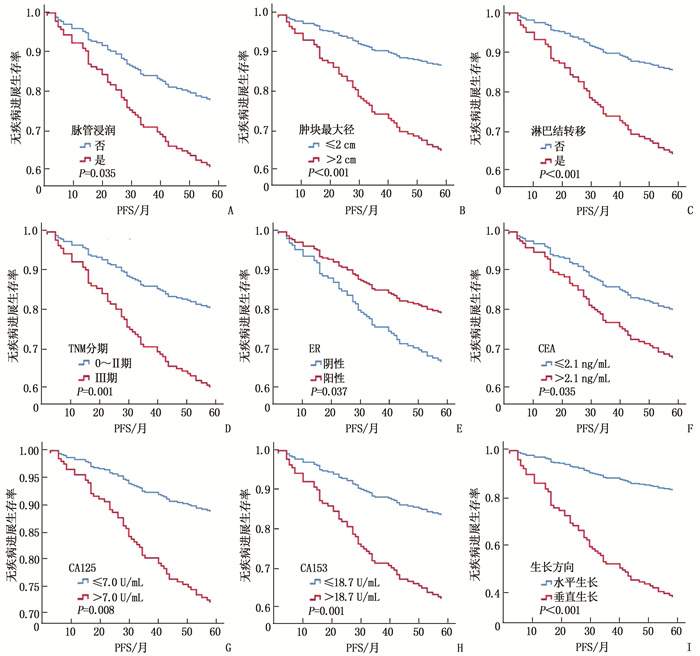
The results of multivariate Cox analysis showed that the largest diameter of tumor, lymph node metastasis, estrogen receptor (ER), carbohydrate antigen 125 (CA125), carbohydrate antigen 153 (CA153) and growth direction were the independent predictors of PFS (P < 0.05). A nomogram model was established based on the above indicators, and the validation results showed that the area under the curve of the receiver operating characteristic (ROC) curve for 5-year PFS was 0.844, and the C-index was 0.793 (95%CI, 0.736 to 0.850), and the 3- and 5-year calibration curves of the nomogram was close to the reference line and showed a good consistency.
In this study, we construct a prognostic prediction nomogram model that combines the clinicopathological features, preoperative cancer markers and ultrasonographic features to provide a visual survival assessment for breast cancer patients.
| [1] |
SIEGEL R L, MILLER K D, FUCHS H E, et al. Cancer Statistics, 2021[J]. CA Cancer J Clin, 2021, 71(1): 7-33. doi: 10.3322/caac.21654
|
| [2] |
赵港美, 章立楠. 乳腺癌患者术后复发转移的危险因素logistic回归分析[J]. 中国妇幼保健, 2021, 36(5): 1147-1149.
|
| [3] |
CIMINO-MATHEWS A. Novel uses of immunohistochemistry in breast pathology: interpretation and pitfalls[J]. Mod Pathol, 2021, 34(suppl 1): 62-77. http://www.nature.com/articles/s41379-020-00697-3
|
| [4] |
GUO Y, HU Y, QIAO M, et al. Radiomics analysis on ultrasound for prediction of biologic behavior in breast invasive ductal carcinoma[J]. Clin Breast Cancer, 2018, 18(3): e335-e344. doi: 10.1016/j.clbc.2017.08.002
|
| [5] |
DI GIOIA D, DRESSE M, MAYR D, et al. Serum HER2 in combination with CA 15-3 as a parameter for prognosis in patients with early breast cancer[J]. Clin Chim Acta, 2015, 440: 16-22. doi: 10.1016/j.cca.2014.11.001
|
| [6] |
MELICHAR B. Laboratory medicine and medical oncology: the tale of two Cinderellas[J]. Clin Chem Lab Med, 2013, 51(1): 99-112. doi: 10.1515/cclm-2012-0496
|
| [7] |
NGUYEN D, YU J, REINHOLD W C, et al. Association of independent prognostic factors and treatment modality with survival and recurrence outcomes in breast cancer[J]. JAMA Netw Open, 2020, 3(7): e207213. doi: 10.1001/jamanetworkopen.2020.7213
|
| [8] |
TANG J N, CUI Q X, ZHANG D, et al. An estrogen receptor (ER)-related signature in predicting prognosis of ER-positive breast cancer following endocrine treatment[J]. J Cell Mol Med, 2019, 23(8): 4980-4990. doi: 10.1111/jcmm.14338
|
| [9] |
SHAO Y, SUN X, HE Y, et al. Elevated levels of serum tumor markers CEA and CA15-3 are prognostic parameters for different molecular subtypes of breast cancer[J]. PLoS One, 2015, 10(7): e0133830. doi: 10.1371/journal.pone.0133830
|
| [10] |
UEHARA M, KINOSHITA T, HOJO T, et al. Long-term prognostic study of carcinoembryonic antigen (CEA) and carbohydrate antigen 15-3(CA 15-3) in breast cancer[J]. Int J Clin Oncol, 2008, 13(5): 447-451. doi: 10.1007/s10147-008-0773-3
|
| [11] |
XU F, LIU F, ZHAO H, et al. Prognostic significance of mucin antigen MUC1 in various human epithelial cancers: a meta-analysis[J]. Medicine: Baltimore, 2015, 94(50): e2286. doi: 10.1097/MD.0000000000002286
|
| [12] |
LI J, LIU L, FENG Z, et al. Tumor markers CA15-3, CA125, CEA and breast cancer survival by molecular subtype: a cohort study[J]. Breast Cancer, 2020, 27(4): 621-630. doi: 10.1007/s12282-020-01058-3
|
| [13] |
ZHANG J Y, WEI Q, DONG D, et al. The role of TPS, CA125, CA15-3 and CEA in prediction of distant metastasis of breast cancer[J]. Clin Chimica Acta, 2021, 523: 19-25. doi: 10.1016/j.cca.2021.08.027
|
| [14] |
LI X, DAI D, CHEN B, et al. Prognostic values of preoperative serum CEA and CA125 levels and nomograms for young breast cancer patients[J]. Onco Targets Ther, 2019, 12: 8789-8800. doi: 10.2147/OTT.S221335
|
| [15] |
REINARTZ S, FAILER S, SCHUELL T, et al. CA125(MUC16) gene silencing suppresses growth properties of ovarian and breast cancer cells[J]. Eur J Cancer, 2012, 48(10): 1558-1569. doi: 10.1016/j.ejca.2011.07.004
|
| [16] |
ZHAO S, MEI Y, WANG J, et al. Different levels of CEA, CA153 and CA125 in milk and benign and malignant nipple discharge[J]. PLoS One, 2016, 11(6): e0157639. doi: 10.1371/journal.pone.0157639
|
| [17] |
朱琳, 郑燕, 薛剑桥, 等. 常规超声联合超声造影对乳腺囊实性复合肿块良恶性的鉴别诊断价值[J]. 中华医学超声杂志: 电子版, 2020, 17(12): 1162-1167. doi: 10.3877/cma.j.issn.1672-6448.2020.12.003
|
| [18] |
PAN B, YAO R, ZHU Q L, et al. Clinicopathological characteristics and long-term prognosis of screening detected non-palpable breast cancer by ultrasound in hospital-based Chinese population (2001-2014)[J]. Oncotarget, 2016, 7(47): 76840-76851. doi: 10.18632/oncotarget.12319
|
| [19] |
CHAE E Y, MOON W K, KIM H H, et al. Association between ultrasound features and the 21-gene recurrence score assays in patients with oestrogen receptor-positive, HER2-negative, invasive breast cancer[J]. PLoS One, 2016, 11(6): e0158461. doi: 10.1371/journal.pone.0158461
|
| [20] |
DIALANI V, GAUR S, MEHTA T S, et al. Prediction of low versus high recurrence scores in estrogen receptor-positive, lymph node-negative invasive breast cancer on the basis of radiologic-pathologic features: comparison with oncotype DX test recurrence scores[J]. Radiology, 2016, 280(2): 370-378. doi: 10.1148/radiol.2016151149
|
| [21] |
WANG H, ZHAN W, CHEN W, et al. Sonography with vertical orientation feature predicts worse disease outcome in triple negative breast cancer[J]. Breast, 2020, 49: 33-40. doi: 10.1016/j.breast.2019.10.006
|
| [22] |
GUO Q, ZHANG L, DI Z, et al. Assessing Risk Category of Breast Cancer by Ultrasound Imaging Characteristics[J]. Ultrasound Med Biol, 2018, 44(4): 815-824. doi: 10.1016/j.ultrasmedbio.2017.12.001
|
| [23] |
杨韵贤, 李世梅, 姚继祎, 等. 美国癌症联合委员会第八版乳腺癌预后分期Ⅰ-Ⅲ期与肿物BI-RADS分类超声特征的关系[J]. 实用医学杂志, 2020, 36(22): 3140-3143. doi: 10.3969/j.issn.1006-5725.2020.22.022
|
| 1. |
朱国玉,吴洋,秦陈,张晓春,李文骥. 中老年Ⅱ~Ⅲ期胃癌患者预后预测列线图模型的构建和验证. 实用临床医药杂志. 2024(17): 27-34+40 .
 本站查看 本站查看
| |
| 2. |
朱婷,周海兰,华骁帆. 基于多维度指标预测乳腺癌术后复发的列线图模型建立及应用. 实用临床医药杂志. 2023(05): 43-48 .
本站查看
| |
| 3. |
林建琴,蒋田华,於晓平,祝娉婷. 乳腺癌患者经外周静脉置入中心静脉导管维护依从性影响因素及精准护理效果分析. 实用临床医药杂志. 2023(16): 142-148 .
本站查看
|
© 2020 《实用临床医药杂志》编辑部
Address: 江苏省扬州市江阳中路136号,扬州大学江阳路北校区14号楼201室China Pos: 225009Tel: 0514-87978917、87978989、87978807
Supported by:
Beijing Renhe Information Technology Co., Ltd.

 苏公网安备 32100302010246号
 DownLoad: