
Value of microcirculation resistance index in evaluating prognosis of patients with acute anterior ST-segment elevation myocardial infarction after emergency percutaneous coronary intervention andrelated influencing factors
-
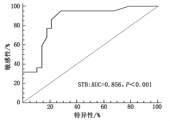
摘要:目的 探讨微循环阻力指数(IMR)评估急性前壁ST段抬高型心肌梗死(STEMI)患者急诊冠状动脉介入治疗(PCI)预后的价值及影响因素。方法 选取行急诊PCI的急性前壁STEMI患者50例, 在PCI后立即测量IMR。以IMR=40 U为临界值,将IMR < 40 U定义为微循环正常组(A组),将IMR≥40 U定义为微循环异常组(B组)。分析2组患者一般资料、实验室检查结果、术中相关情况,以及术后24 h、术后6个月、术后12个月的超声心动图结果及术后随访12个月的主要心脏不良事件(MACE)情况。结果 Logistic回归分析发现,症状发作至球囊扩张时间(STB)是影响急性前壁STEMI患者微循环障碍的独立危险因素。在PCI术后24 h、6个月、12个月时, 2组左室射血分数(LVEF)、左室舒张末容积内径(LVEDD)比较,差异有统计学意义(P < 0.05)。2组患者MACE情况比较,差异有统计学意义(P < 0.05)。结论 STB是急性前壁STEMI患者微循环障碍的独立危险因素, IMR对急性前壁STEMI患者PCI术后左心重构、左心功能恢复及主要心脏不良事件的发生可能有较好的早期预测价值。
-
关键词:
- 急性ST段抬高型心肌梗死 /
- 微循环阻力指数 /
- 微循环障碍 /
- 急诊经皮冠状动脉介入治疗 /
- 心脏不良事件 /
- 左室射血分数
Abstract:Objective To investigate the value of microcirculation resistance index (IMR) in evaluating prognosis of patients with acute anterior ST-segment elevation myocardial infarction (STEMI) after emergency percutaneous coronary intervention (PCI) and related influencing factors.Methods Totally 50 patients with acute anterior STEMI by PCI were selected, and IMR was measured immediately after PCI. Taking IMR=40 U as the cutoff value, patients with IMR < 40 U was defined as normal microcirculation group (group A), and those with IMR≥40 U was defined as abnormal microcirculation group (group B). The general data, laboratory results, intraoperative information, and echocardiographic results at 24 hours, 6 months and 12 months after operation were analyzed. Major adverse cardiac events (MACE) at 12 months after operation were analyzed.Results Logistic regression analysis showed that time from onset of symptoms to balloon dilation (STB) was an independent risk factor for microcirculation disorders in acute anterior STEMI patients. At 24 hours, 6 months and 12 months after PCI, there were significant differences in the left ventricular ejection fraction (LVEF) and left ventricular end-diastolic volume diameter (LVEDD) between the two groups (P < 0.05). There was a significant difference in MACE between the two groups (P < 0.05).Conclusion STB is an independent risk factor for microcirculation disorders in acute anterior STEMI patients. IMR may have a good early predictive value for left ventricular remodeling, recovery of left cardiac function and incidence of MACE after PCI in acute anterior STEMI patients. -
-
表 1 2组患者一般资料比较(x±s)[n(%)]
指标 A组(n=28) B组(n=22) 年龄/岁 <65 21(75.00) 9(40.91)* ≥65 7(25.00) 13(59.09)* 心率/(次/min) 76.50±14.74 76.05±14.69 收缩压/mmHg 135.96±20.46 125.73±20.09 舒张压/mmHg 82.96±12.95 80.78±12.69 性别 男 25(89.29) 18(81.82) 女 3(10.71) 4(18.18) 糖尿病史 无 17(60.71) 16(72.73) 有 11(39.29) 6(27.27) 高血压病史 无 9(32.14) 11(50.00) 有 19(67.86) 11(50.00) 吸烟史 无 9(32.14) 5(22.73) 有 19(67.86) 17(77.27) 高脂血症病史 无 23(82.14) 16(72.73) 有 5(17.86) 6(27.27) 与A组比较, *P < 0.05。  下载: 导出CSV
表 2 2组患者实验室检查结果比较(x±s)
指标 A组(n=28) B组(n=22) HGB/(g/L) 147.00±17.29 138.05±21.02 PLT/(×109/L) 203.39±66.69 181.23±42.08 CREA/(μmol/L) 72.71±18.81 67.45±14.61 LDL-C/(mmol/L) 2.84±0.85 2.60±0.93 尿酸/(mmol/L) 335.57±82.99 340.27±91.44 cTnI/(ng/mL) 7.75(1.25, 24.90) 10.98(2.19, 26.54) CK-MB峰值/(ng/mL) 12.90(4.00, 48.09) 44.08(13.65, 72.75)* MYO/(ng/mL) 124.60(75.12, 298.48) 298.21(75.89, 637.89) D-D/(mg/L) 0.26(0.17, 0.42) 0.35(0.18, 0.60) NLR 4.6(2.52, 7.55) 8.69(5.42, 12.31)* HGB: 血红蛋白; PLT: 血小板计数; CREA: 肌酐;
LDL-C: 低密度脂蛋白胆固醇; cTnI: 肌酐蛋白I;
CK-MB: 肌酸激酶同工酶; MYO: 肌红蛋白; D-D: D-二聚体;
NLR: 中性粒细胞计数与淋巴细胞计数比值。与A组比较, *P < 0.05。
下载: 导出CSV
表 3 2组患者术中情况比较[n(%)]
指标 A组(n=28) B组(n=22) 血栓抽吸 否 22(78.57) 16(72.73) 是 6(21.43) 6(27.27) 球囊后扩 否 16(57.14) 9(40.91) 有 12(47.14) 13(59.09) Ⅱb/Ⅲa受体拮抗剂 无 12(47.14) 8(36.36) 替罗非班 16(57.14) 14(63.64) 硝普钠 否 24(85.71) 17(77.27) 有 4(14.29) 5(22.73) 合并其他冠脉病变 合并回旋支病变 14(50.00) 7(31.82) 合并右冠病变 11(39.29) 8(36.36) 同时合并回旋支及右冠病变 9(32.14) 6(27.27) 支架长/mm 25(23.25, 32.25) 30(20.00, 38.25) 支架直径/mm 3(3.00, 3.50) 3(3.00, 3.50) STB/min 240(180.00, 300.00) 383(332.25, 525.00)* STB: 症状发作至球囊扩张时间; NLR: 中性粒细胞计数与淋巴细胞计数比值。与A组比较, *P < 0.05。
下载: 导出CSV
表 4 急性前壁STEMI患者发生微循环障碍的危险因素
因素 B SE P OR 95%CI 下限 上限 年龄≥65岁 1.359 0.864 0.116 3.894 0.716 21.178 CK-MB峰值 0.006 0.006 0.307 1.006 0.995 1.017 NLR 0.206 0.123 0.093 1.229 0.966 1.563 STB 0.012 0.004 0.003 1.012 1.004 1.02 CK-MB: 肌酸激酶同工酶; STB: 症状发作至球囊扩张时间; NLR: 中性粒细胞计数与淋巴细胞计数比值。
下载: 导出CSV
表 5 2组患者术后左心功能比较
组别 时点 LVEF/% LVEDD/mm A组(n=28) 术后24 h 50.79±7.198 47.46±4.17 术后6个月 55.00±6.837# 47.43±3.75 术后12个月 56.32±6.700# 49.61±4.18#▲ B组(n=22) 术后24 h 43.59±6.284* 51.09±4.79* 术后6个月 49.41±10.477*# 51.14±5.83* 术后12个月 42.05±8.677*▲ 53.77±5.31*#▲ LVEF: 左室射血分数; LVEDD: 左室舒张末容积内径。
与A组相比, *P < 0.05; 与术后24 h相比, #P < 0.05;
与术后6个月相比, ▲P < 0.05。
下载: 导出CSV
-
[1] De Maria G L, Cuculi F, Patel N, et al. How does coronary stent implantation impact on the status of the microcirculation during primary percutaneous coronary intervention in patients with ST-elevation myocardial infarction[J]. European Heart Journal, 2015, 36(45): 3165-3177. doi: 10.1093/eurheartj/ehv353
[2] Cuculi F, De Maria G L, Meier P, et al. Impact of Microvascular Obstruction on the Assessment of Coronary Flow Reserve, Index of Microcirculatory Resistance, and Fractional Flow Reserve After ST-Segment Elevation Myocardial Infarction[J]. Journal of the American College of Cardiology, 2014, 64(18): 1894-1904. doi: 10.1016/j.jacc.2014.07.987
[3] Fiarresga A, Selas M, Oliveira E, et al. Invasive assessment of the coronary microcirculation using the index of microcirculatory resistance: Description and validation of an animal model[J]. Rev Port Cardiol, 2014, 34(4): 207-212. http://www.sciencedirect.com/science/article/pii/S217420491400097X
[4] Bonello L, Ait M O, Lemesle G, et al. Incidence and predictors of microvascular dysfunction assessed by the index of microcirculatory resistance following primary PCI for ST-elevation myocardial infarction[J]. Int J Cardiol, 2011, 146(3): 465-467. doi: 10.1016/j.ijcard.2010.10.134
[5] Ng M K C, Yeung A C, Fearon W F. Invasive Assessment of the Coronary Microcirculation Superior Reproducibility and Less Hemodynamic Dependence of Index of Microcirculatory Resistance Compared With Coronary Flow Reserve[J]. Circulation, 2006, 113(17): 2054-2061. doi: 10.1161/CIRCULATIONAHA.105.603522
[6] Faustino M, Baptista S B, Freitas A, et al. The Index of Microcirculatory Resistance as a Predictor of Echocardiographic Left Ventricular Performance Recovery in Patients With ST-Elevation Acute Myocardial Infarction Undergoing Successful Primary Angioplasty[J]. Journal of Interventional Cardiology, 2016, 29(2): 137-145. doi: 10.1111/joic.12278
[7] Park S, Baek Y, Lee M, et al. Comprehensive assessment of microcirculation after primary percutaneous intervention in ST-segment elevation myocardial infarction: insight from thermodilution-derived index of microcirculatory resistance and coronary flow reserve[J]. Coronary Artery Disease, 2016, 27(1): 34-39. doi: 10.1097/MCA.0000000000000310
[8] 王世超, 刘玉昊, 武越, 等. 微循环阻力指数对冠状动脉完全闭塞病变择期行介入治疗的预测价值及影响因素[J]. 中国循环杂志, 2016(4): 332-336. doi: 10.3969/j.issn.1000-3614.2016.04.005 [9] Park S, Baek Y, Lee M, et al. Comprehensive assessment of microcirculation after primary percutaneous intervention in ST-segment elevation myocardial infarction[J]. Coronary Artery Disease, 2016, 27(1): 34-39. doi: 10.1097/MCA.0000000000000310
[10] Ahmed N, Layland J, Carrick D, et al. Safety of guidewire-based measurement of fractional flow reserve and the index of microvascular resistance using intravenous adenosine in patients with acute or recent myocardial infarction[J]. International Journal of Cardiology, 2016, 202: 305-310. doi: 10.1016/j.ijcard.2015.09.014
[11] De Maria G L, Alkhalil M, Wolfrum M, et al. Index of microcirculatory resistance as a tool to characterize microvascular obstruction and to predict infarct size regression in patients with STEMI undergoing primary PCI[J]. JACC Cardiovasc Imaging, 2019, 12(5): 837-848. doi: 10.1016/j.jcmg.2018.02.018
[12] Fearon W F, Low A F, Yong A S, et al. Prognostic Value of the Index of Microcirculatory Resistance Measured After Primary Percutaneous Coronary Intervention[J]. Circulation, 2013, 127(24): 2436-2441. doi: 10.1161/CIRCULATIONAHA.112.000298
[13] Palmer S, Layland J, Carrick D, et al. The Index of Microcirculatory Resistance Postpercutaneous Coronary Intervention Predicts Left Ventricular Recovery in Patients With Thrombolyzed ST-Segment Elevation Myocardial Infarction[J]. Journal of Interventional Cardiology, 2016, 29(2): 146-154. doi: 10.1111/joic.12271
[14] Baek Y, Park S, Kim S, et al. Clinical and Angiographic Predictors of Microvascular Dysfunction in ST-Segment Elevation Myocardial Infarction[J]. Yonsei Medical Journal, 2015, 56(5): 1235-1239. doi: 10.3349/ymj.2015.56.5.1235
[15] Lee M, Park S, Kwon S W, et al. Relation Between Neutrophil-to-Lymphocyte Ratio and Index of Microcirculatory Resistance in Patients With ST-Segment Elevation Myocardial Infarction Undergoing Primary Percutaneous Coronary Intervention[J]. The American Journal of Cardiology, 2016, 118(9): 1323-1328. doi: 10.1016/j.amjcard.2016.07.072
[16] Pan W, Zhao D, Zhang C, et al. Application of neutrophil/lymphocyte ratio in predicting coronary blood flow and mortality in patients with ST-elevation myocardial infarction undergoing percutaneous coronary intervention[J]. Journal of Cardiology, 2015, 66(1): 9-14. doi: 10.1016/j.jjcc.2014.10.014
[17] Park J J, Jang H, Oh I, et al. Prognostic Value of Neutrophil to Lymphocyte Ratio in Patients Presenting With ST-Elevation Myocardial Infarction Undergoing Primary Percutaneous Coronary Intervention[J]. The American Journal of Cardiology, 2013, 111(5): 636-642. doi: 10.1016/j.amjcard.2012.11.012
[18] Fearon W F, Shah M, Ng M, et al. Predictive Value of the Index of Microcirculatory Resistance in Patients With ST-Segment Elevation Myocardial Infarction[J]. Journal of the American College of Cardiology, 2008, 51(5): 560-565. doi: 10.1016/j.jacc.2007.08.062
[19] Sabin P, Koshy A G, Gupta P N, et al. Predictors of no- reflow during primary angioplasty for acute myocardial infarction, from Medical College Hospital, Trivandrum[J]. Indian Heart Journal, 2017, 69: S34-S45. http://www.sciencedirect.com/science/article/pii/S0019483216304205
[20] 魏腾飞, 赵蓓, 刘佩林, 等. 发病至首次医疗接触时间对急性ST段抬高型心肌梗死患者预后影响的研究[J]. 中华心血管病杂志, 2017, 45(5): 393-398. doi: 10.3760/cma.j.issn.0253-3758.2017.05.006 [21] Park S, Baek Y, Lee M, et al. Comprehensive assessment of microcirculation after primary percutaneous intervention in ST-segment elevation myocardial infarction[J]. Coronary Artery Disease, 2016, 27(1): 34-39. doi: 10.1097/MCA.0000000000000310
-
期刊类型引用(12)
1. 苗雪艳,彭韶,储卫红. 肠道菌群失调与RRTI患儿T淋巴细胞亚群、免疫球蛋白水平的关系. 海南医学. 2024(17): 2501-2505 .  百度学术
百度学术
2. 刘军,陈秋萍,毛晓玲,刘明. 探讨脾氨肽在儿童呼吸道反复感染不同类型中的作用. 药品评价. 2021(11): 695-697 . 百度学术
3. 邓春荣,姜静,任秀,张凯,于代华,范洪伟. 脾多肽对ICU呼吸机相关性肺炎患者细胞免疫功能和治疗效果的影响. 重庆医学. 2020(08): 1274-1278 . 百度学术
4. 朱长龙,韦红,马晓燕. 反复呼吸道感染患儿血清维生素A、E水平与T细胞免疫的关系研究. 中国儿童保健杂志. 2020(07): 798-801 . 百度学术
5. 崔洁媛,李春珍,张东风,袁小颖,刘玲,刘福娟. 脾多肽注射液辅助治疗儿童紫癜性肾炎的疗效. 中华临床医师杂志(电子版). 2020(08): 630-634 . 百度学术
6. 刘海芬. 阿奇霉素治疗儿童呼吸道感染的治疗效果. 世界最新医学信息文摘. 2019(15): 154+160 . 百度学术
7. 周寒丽,段方方,孔天东. 脾多肽联合化疗治疗对肺癌患者临床疗效和免疫功能的影响. 四川解剖学杂志. 2019(01): 86-87 . 百度学术
8. 李进,方代华. T淋巴细胞亚群与NK细胞在小儿支气管肺炎中的变化及意义. 广西医科大学学报. 2019(05): 785-788 . 百度学术
9. 唐庆,李少宁,詹文娟. 复可托治疗小儿反复肺炎支原体感染的疗效及对免疫功能和肺功能的影响. 广西医科大学学报. 2018(01): 33-37 . 百度学术
10. 崔卫国,崔圣莹. 脾多肽辅助甘草锌颗粒对慢性腹泻小儿血锌水平及免疫功能的影响. 泰山医学院学报. 2018(11): 1238-1240 . 百度学术
11. 刘靖,余南生,谢军. 脾多肽注射液联合芪黄益肺合剂治疗重症肺炎的疗效及对免疫功能的影响. 广东医学. 2018(21): 3266-3269 . 百度学术
12. 张磊,王耀邦,高凤,曹兴丽,侍响响. 阿奇霉素与免疫刺激剂脾多肽联合治疗支原体肺炎的疗效及对细胞免疫水平的影响. 中国妇幼保健. 2017(24): 6168-6171 . 百度学术
其他类型引用(1)
计量
- 文章访问数: 299
- HTML全文浏览量: 141
- PDF下载量: 6
- 被引次数: 13
 苏公网安备 32100302010246号